Fig 6.
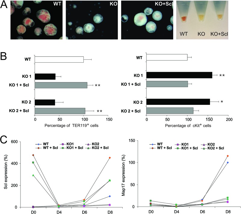
Rescue of the SclΔ40/Δ40 ES cells phenotype by Flag-Scl. (A) Representative photographs of EBs generated from SclWT/WT (WT), SclΔ40/Δ40 (KO), and SclΔ40/Δ40 mice overexpressing Flag-Scl (KO + Scl) and pelleted EBs (right). (B) Histograms showing the percentages of Ter119-positive (left) and cKit-positive (right) cells in day 8 EBs. *, P ≤ 0.05; **, P ≤ 0.01 (compared to KO mice). (C) Expression of Scl (left) and Map17 (right) during embryoid body differentiation. Data are normalized to the expression at day 8 in SclWT/WT EBs. WT, SclWT/WT; KO, SclΔ40/Δ40.

