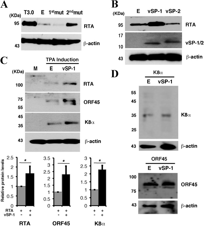
Fig 3.
vSP-1 enhances the expression of RTA. (A) 293T cells were cotransfected with pCR3.1-ORF50 and T3.0 mutant constructs as indicated, followed by Western blotting for the RTA expression level. T3.0-1st mut is T3.0 with a point mutation at position 74356, and T3.0-2nd mut is T3.0 with a point mutation at position 74028 (as diagramed in Fig. 1A). Lane E, empty vector. (B) 293T cells were cotransfected with pCR3.1-ORF50 and either vSP-1, vSP-2, or an empty vector. Then the expression of RTA and vSP-1/2 was analyzed by Western blotting. (C) Effect of vSP-1 on the expression of the KSHV lytic genes downstream of RTA in the RTA-initiated lytic gene expression cascade. BCBL-1 cells were transfected with the vSP-1 expression vector. Twenty-four hours posttransfection, cells were induced by TPA for 16 h. (Top) Cell lysates were subjected to Western blotting for the expression levels of the different KSHV viral proteins by using specific antibodies. (Bottom) The band intensities are plotted graphically. Data are means and standard deviations for three experimental replicates. *, P ≤ 0.05. (D) A Flag-vSP-1 or empty vector was introduced into 293T cells with either a K8α or an ORF45 expression vector. Transfected cells were lysed and were analyzed by Western blotting using antibodies against K8α, ORF45, and β-actin.

