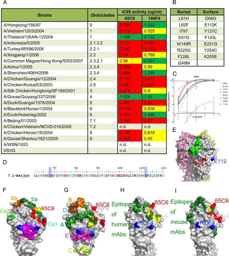
Fig 1.
Amino acid residues involved in the neutralization epitope of 100F4. (A) Broad neutralization activity (95% inhibitory concentration [IC95]) of antibodies 65C6 and 100F4 against a panel of H5N1 pseudotypes reproduced from reference 1. Green, >1 μg/ml required to reach IC95; yellow, between 0.5 and 1 μg/ml required to reach IC95; red, <0.5 μg/ml required to reach IC95. (B) List of 15 single amino acid mutants that antibody 100F4 can no longer bind to, obtained by using yeast Saccharomyces cerevisiae that displays a random mutagenesis library of HA fragment comprising amino acid residues 51 to 260 in fine epitope mapping. Among them, 8 amino acid mutants are underneath the HA surface and the other 7 are on the surface of HA. (C) Titration of antibody 100F4 against H5N1 pseudotypes expressing 7 single surface amino acid mutants compared to the results against H5N1 pseudotype expressing the parental HA. (D) Amino acid residues D68 and E112 (D72 and E116 in H3 numbering) involved in 100F4 epitope are highlighted by blue shading. (E) Amino acid residues D68 and E112 are highlighted in red and blue, respectively, in space-filling model of HA. Yellow, pink, and grey each indicate one of three monomers that make up an HA trimer. (F) 100F4 and 65C6 epitopes in the context of known neutralization epitopes in the H1 HA structure (Protein Data Bank [PDB] structure accession number IRU7), as follows: Ca1, cyan; Ca2, light green; Cb, magenta; Sa, forest green; and Sb, orange. Yellow, overlap amino acid residues between the 65C6 epitope and the Sa site. (G) 100F4 and 65C6 epitopes in the context of known neutralization epitopes in H3 HA structure (PBD structure accession number 2VIU), as follows: site A, forest green; site B, orange; site C, yellow-green; site D, cyan; and site E, magenta. Yellow, overlap amino acid residues between the 65C6 epitope and site A. (H) 100F4 and 65C6 epitopes in the context of known neutralization epitopes detected by human MAb in H5 HA structure (PBD structure accession number 2ibx). Neutralization epitopes detected by human MAb are highlighted in forest green, and amino acid residues that overlap amino acid residues of the 65C6 epitope are highlighted in yellow. (I) 100F4 and 65C6 epitopes in the context of known neutralization epitopes detected by mouse MAb in H5 HA structure (PBD structure accession number 2ibx). Neutralization epitopes detected by mouse MAb are highlighted in forest green, and amino acid residues that overlap amino acid residues of 65C6 epitope are highlighted in yellow.

