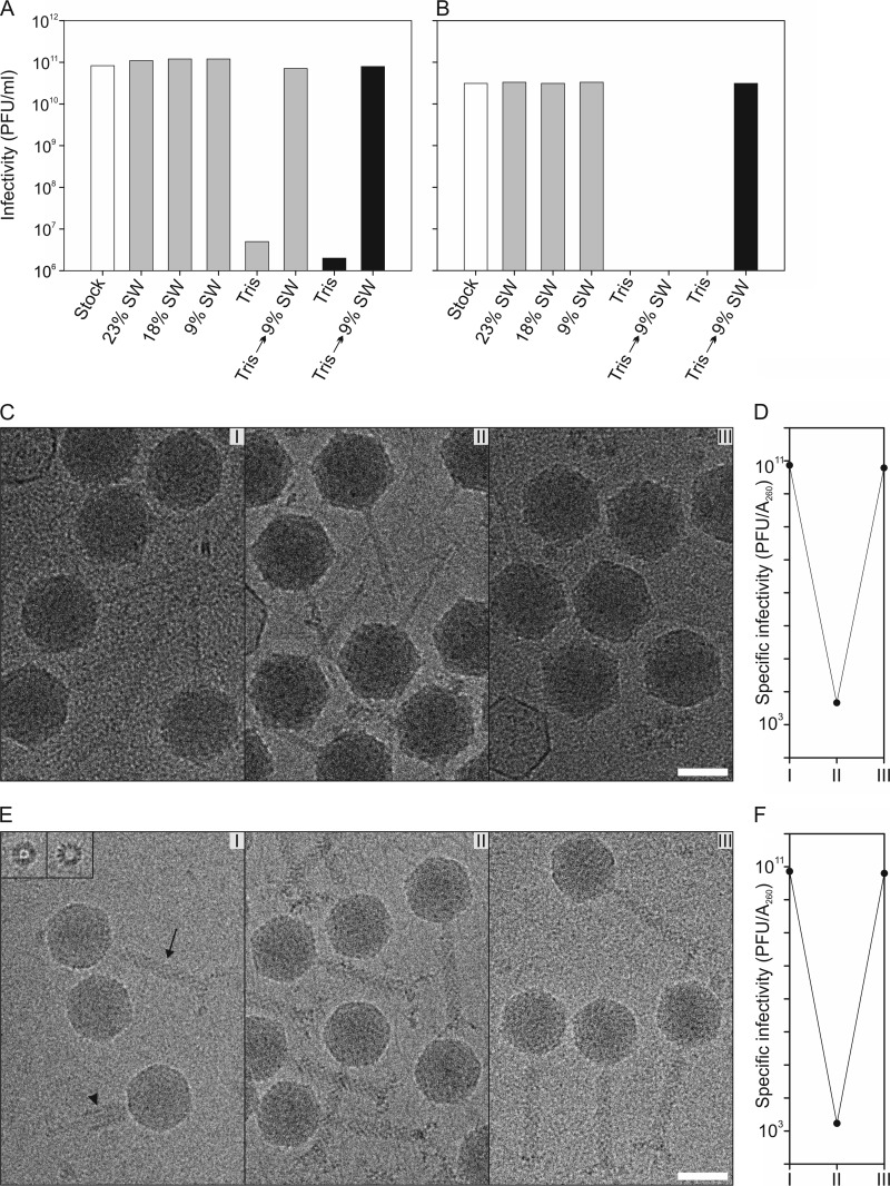
Fig 2.
Viruses are reversibly inactivated under conditions of low salinity without observable morphological changes. (A and B) Effect of lowered ionic strength on the infectivity of HVTV-1 (A) and HSTV-2 (B). The virus stocks (white bars) were diluted either 100-fold (black bars) or 1,000-fold (gray bars) in 23, 18, or 9% salt water (SW) or in 20 mM Tris-HCl (pH 7.2) and incubated for 1 h at 4°C. After incubation, the samples were further diluted in the same buffer, except that the Tris-HCl samples were diluted in 9% SW as well. The diluted samples were incubated for 15 min at RT, and infectivity was then determined. The bars represent the averages of data from two experiments. The infectivity scale in panels A and B is the same, and the infectivities are corrected for the dilution. (C) Cryo-electron micrographs of HVTV-1 at a 4-μm underfocus. (I) Virions incubated in 9% SW; (II) virions incubated in 20 mM Tris-HCl (pH 7.2) buffer; (III) virions which were first incubated in 20 mM Tris-HCl (pH 7.2) and then restored in 9% SW. Bar, 50 nm. (D) Specific infectivity on a logarithmic scale of HVTV-1 virions at different salinities (I to III). (E) Cryo-electron micrographs of HSTV-2 at a 4-μm underfocus showing particles as in panel A. In panel I, the arrow indicates a noncontracted tail, the arrowhead indicates a contracted tail, and the insets show longitudinal views of baseplates dissociated from the capsids. Bar, 50 nm. (F) Specific infectivity on a logarithmic scale of HSTV-2 virions at different salinities (I to III).

