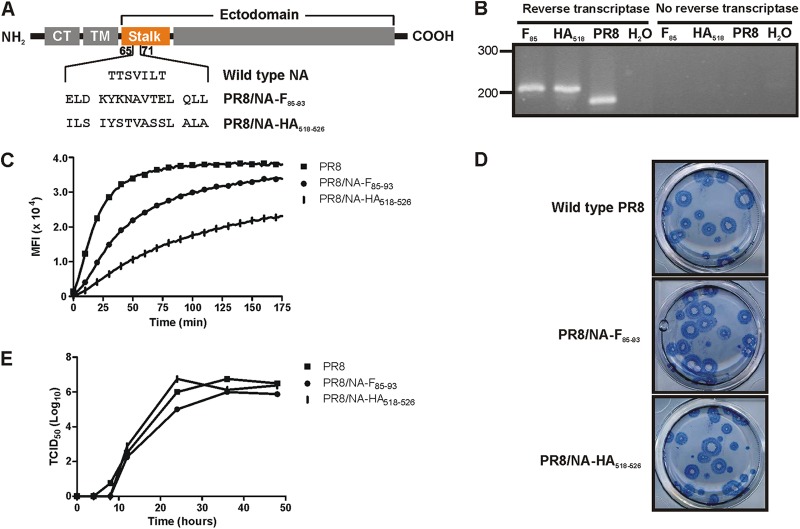
Fig 1.
Characterization of recombinant PR8/NA-F85–93 and PR8/NA-HA518–526 influenza viruses. (A) Schematic representation of wild-type and mutant neuraminidases. CT, cytoplasmic tail; TM, transmembrane domain. (B) RT-PCR of an NA fragment containing the insertion site of the epitope. A band shift from 170 to 207 bp was seen when the epitope was inserted. (C) Neuraminidase activity assay on live, purified virus. Fluorescence of the cleaved MUNANA substrate was measured every 2 min during 3 h. (D) Plaque phenotypes of the PR8/NA-F85–93 and PR8/NA-HA518–526 influenza viruses do not differ from the wild-type virus plaques in an MDCK plaque assay. (E) In vitro growth kinetics. MDCK cells were infected at a multiplicity of infection of 0.001 of wild-type PR8, PR8/NA-F85–93, or PR8/NA-HA518–526 virus. Samples were taken at 0, 4, 8, 12, 24, and 48 h postinfection. The viral titer in the samples was determined by a TCID50 assay.

