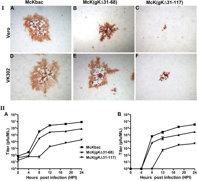
Fig 2.
Plaque morphology and replication kinetics of the McKbac and gK mutant viruses. (I) Plaque morphology. Vero cells were infected with each virus at an MOI of 0.001, and at 48 hpi viral plaques were fixed with methanol and stained with anti-HSV antibodies as described in Materials and Methods. Representative viral plaques of all gK mutant viruses and the McKbac virus are shown on both Vero (A to C) and VK302 (D to F) cells. (II) Replication kinetics. Vero cells were infected with each virus at either a low MOI (0.1) or a high MOI (2), and the numbers of infectious viruses produced were determined on VK302 cells at different times postinfection. Viral titers after high-MOI infection are shown in panel A, and low-MOI infections are shown in panel B. All experiments were performed in triplicate. Error bars represent standard errors of the means.

