
Fig 7.
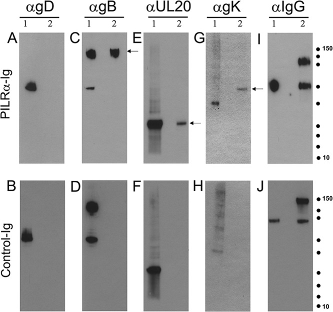
Soluble PILRα interacts with gB, gK, and UL20p in virus-infected cells. Western blot analysis of immunoprecipitates of PILRα ligand from HSV-1-infected cells is shown. Vero cells were infected with a double-tagged HSV-1 virus (YE102-VC1). (A to H) Infected lysates were immunoprecipitated with PILRα-Ig or CD200-Ig (control) and the immunoprecipitates were separated by SDS-PAGE, followed by blotting with anti-gB, anti-gD, anti-V5 (gK), and anti-FLAG (UL20) Ab. (I and J) The Ig fusion proteins used for immunoprecipitation were detected by anti-human IgG Ab. Molecular mass standards are shown as dots (150, 100, 75, 50, 37,25, 20, 15, and 10 kDa; Precision Plus protein standards; Bio-Rad). Lane 1, lysate from infected cells; lane 2, lysates immunoprecipitated with PILRα-Ig or CD200-Ig. Arrows indicate protein species of interest.