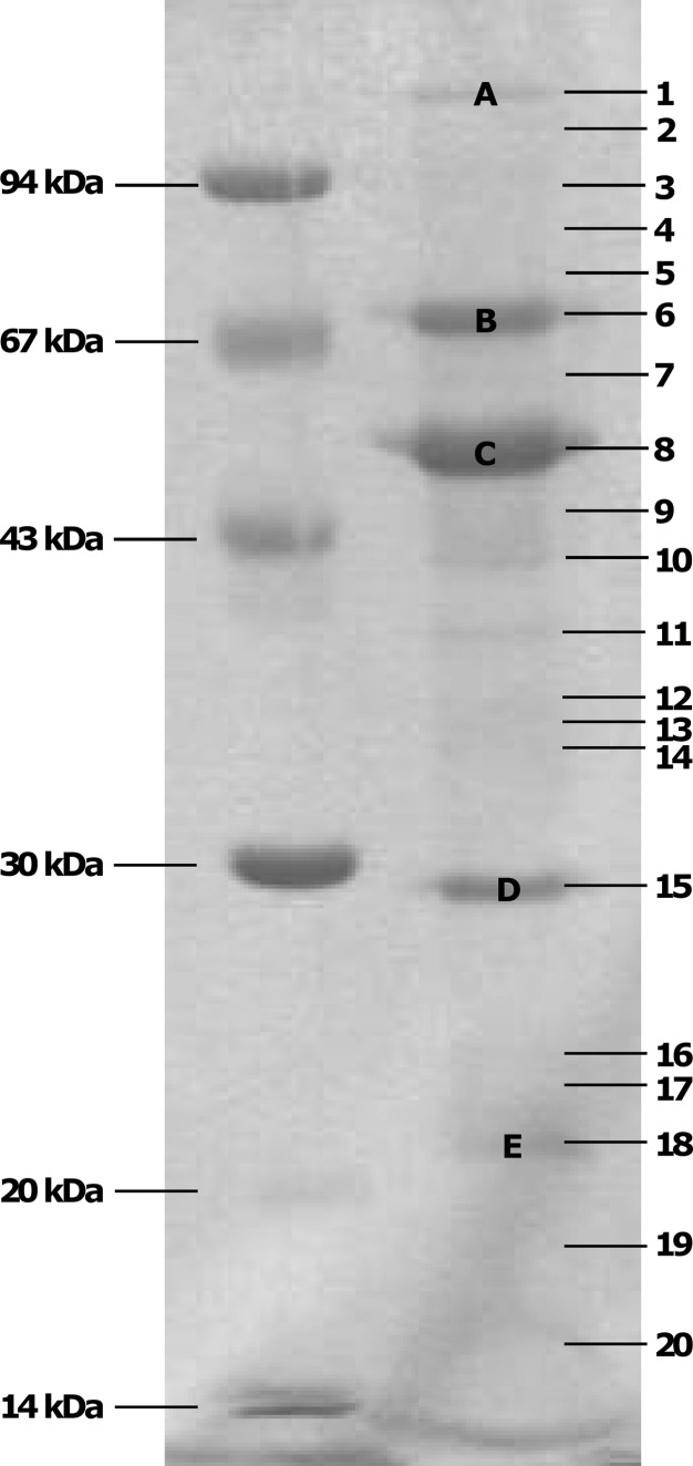
Fig 5.
Coomassie G-250-stained SDS-PAGE gel of the structural proteome of bacteriophage Remus. A low-molecular-mass marker is indicated on the left. The numbers on the right correspond to the analyzed gel pieces of which the identified proteins are listed in Table 2. Prominent protein bands are indicated with capital letters.

