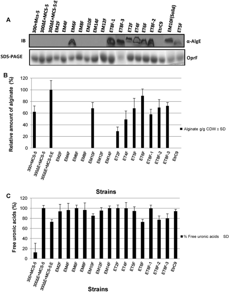
Fig 4.
Localization of alginate and free uronic acid production by FLAG epitope insertion variants of AlgE. (A) The OM fraction isolated from planktonic cultures (unless mentioned from solid medium) of P. aeruginosa PDO300ΔalgE harboring various plasmids, showing the absence or presence of AlgE and its variants. The presence of AlgE and its variants in the OM can be seen in the immunoblots (IB) probed with anti-AlgE antibodies (upper panel). EM10F(Solid) indicates that the OM was isolated from PDO300ΔalgE (MCS5::algEM10F) cells grown on solid medium. Constitutively expressed oprF was used as a loading control (bottom panel). Only the relevant parts of the different gels and the immunoblots are shown. The bands shown here are not from a single blot. Some bands were pasted in after cutting from a different blot. (B) The amount of alginate produced was assessed by growing cells on solid medium, and results are presented relative to the amount of alginate produced by P. aeruginosa PDO300ΔalgE (pBBRMCS-5::algE). (C) The amount of free uronic acid was assessed after overnight growth in liquid culture and is given as the percent ratio between the filtrate and the supernatant. Experiments were conducted in triplicate. Error bars show the standard deviations of the mean values. 300+MCS-5 indicates PDO300 carrying the pBBR1MCS-5 plasmid; 300ΔE+MCS-5 shows PDO300ΔalgE carrying the pBBR1MCS-5 plasmid; 300ΔE+MCS-5:E depicts PDO300ΔalgE carrying the pBBR1MCS-5::algE plasmid. Variants of AlgE are indicated with the first letter E, indicating wild-type AlgE, followed by M for the membrane segment or T for the periplasmic turn, followed by a number that indicates the position in AlgE as defined by the structure shown schematically in Fig. 1. The letter F is used as an abbreviation for the FLAG epitope.

