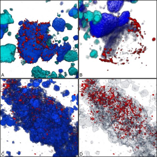
Fig 4.
Three-dimensional visualization showing the coaggregation of the Micavibrio-like bacterium with sublineage I Nitrospira. (A) Micavibrio-like cells (red) attached to a cell aggregate of sublineage I Nitrospira (blue). Other nitrifiers (AOB and sublineage II Nitrospira) are shown in cyan. Applied probes were HSAL723 and HSAL866 (both labeled with Cy3), Ntspa662 (labeled with Cy5, rendered as blue), Ntspa1151 (labeled with FLUOS, rendered as cyan), and NEU, Cluster6a192, and Nso1225 (labeled with FLUOS, rendered as cyan). (B) Micavibrio-like cells surrounding a partly dark (possibly degraded) sublineage I Nitrospira cell aggregate. Colors and probes are like in panel A. (C) Micavibrio-like cells (red) attached to a cell aggregate of Nitrospira (probe Ntspa662; blue). (D) Same position as shown in panel C, but Nitrospira clusters are rendered semitransparent to show that Micavibrio-like cells also penetrated the inner parts of the Nitrospira cell colonies.

