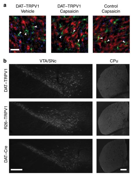
Figure 2. Capsaicin administration does not induce apparent cell death in DAT–TRPV1 mice.
(a) Pseudo-coloured images of fluorescent immunohistochemistry with TH (red), ionized calcium-binding molecule-1 (Iba-1, a marker for microglial cells; green) antibody and DAPI (blue) staining 24 h after capsaicin (32 mg kg −1) or vehicle administration. microglia (arrowheads) remain ramified in the midbrain of DAT–TRPV1 mice following capsaicin administration implicating normal homeostasis and lack of neuropathology. scale bar, 50 μm. (b) Following 10 i.p. injections of capsaicin (32 mg kg −1) through 30-day period, TH fluorescent immunohistochemistry of VTA/snc and CPu of DAT–TRPV1, R26-TRPV1 or DAT-Cre mice (n = 3–4 per group). only a single dose of capsaicin was administered per day. scale bars, 200 μm.

