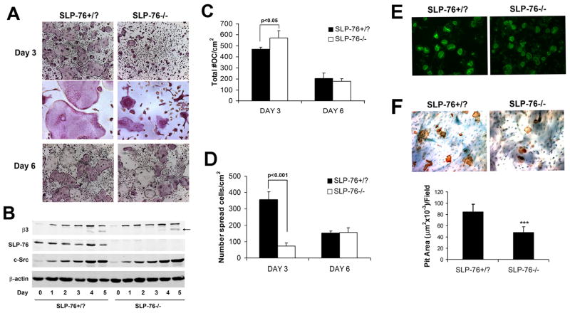
Figure 2. SLP-76-deficient BMMs differentiate into OCs normally but exhibit delayed spreading and reduced resorptive capacity.
(A) SLP-76+/? and SLP-76−/− BMMs, isolated from radiation chimera marrow, were cultured in M-CSF and RANKL for 3 or 6 days. The cells were stained for TRAP activity (top and bottom panels, magnification 40x, middle panel 200x). (B) SLP-76+/? and SLP-76−/− BMMs were cultured in M-CSF and RANKL, with time. Lysates were immunoblotted for the OC differentiation markers, β3 integrin subunit and c-Src; β-actin serves as a loading control. (C, D) Quantification of C) total and D) spread OCs in (A). (E) SLP-76+/− and SLP-76−/− BMMs were cultured on bone slices, with M-CSF and RANKL, for six days. The resultant OCs were labeled with FITC-phalloidin to visualize actin rings (top panel) (200x). They were then removed and resorptive pits stained with peroxidase-conjugated WGA followed by 3-3′-diaminobenzidine (bottom panel) (200x). (F) Quantification of resorptive pit area in (D). (***p<0.001)

