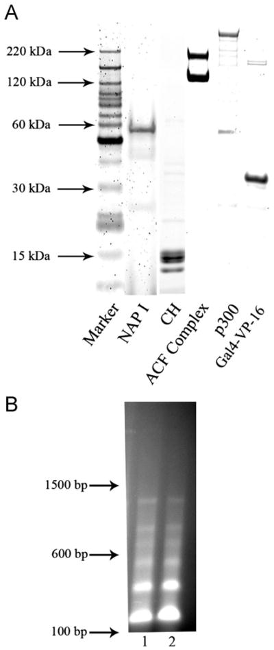
Fig. 1.
(A) SDS-PAGE gel of the purified protein components required for the assembly and in vitro transcription of chromatin templates. Proteins were purified as described in Materials and methods. Aliquots were subjected to SDS-PAGE electrophoresis and the protein bands stained with SYPRO orange and visualized by laser fluoroscopy as described. Proteins preparations were of the expected size. (B) Micrococal nuclease digests of a representative pG5MLT reconstituted chromatin template shows the expected regularly spaced nucleosome pattern. Lanes 1 and 2 are 150 ng of pG5MLT reconstituted chromatin digested with 1.8 units of micrococcal nuclease for 3 and 9 min respectively. Indicated sizes reflect the migration of a 100 bp DNA ladder.

