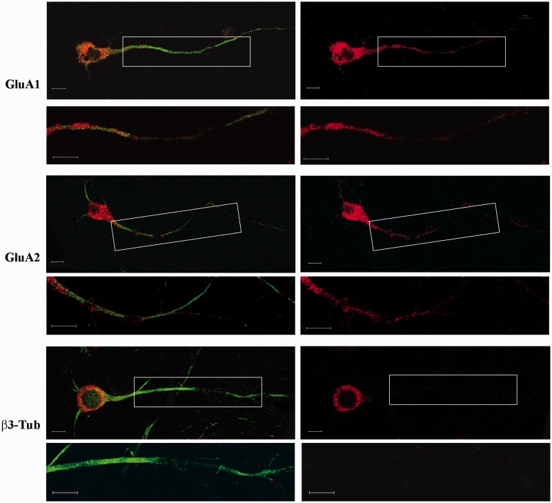
Figure 1.
Fluorescence in situ hybridization of GluA1, GluA2 and β3-Tub mRNAs in primary cortical neurons. The right panel shows the hybridization signal (red) of the different mRNAs. In the left panel, the figures are merged with the immunostaining for MAP2. Under each panel, high magnification of the boxed dendritic regions are reported. Scale bar is 10 µm.

