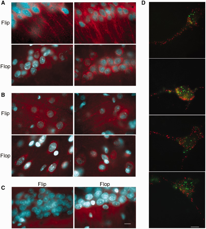
Figure 6.
Fluorescent in situ hybridization of GluA2 mRNA in Flip and Flop splicing variants in (A) CA1 hippocampal and (B) cortical coronal sections. (C) The sense probe was used as negative control. Flip Flop hybridizations were performed on consecutive sections. (D) In situ detection of individual transcripts with padlock probes and target-primed RCA. After in situ reverse transcription and RNA degradation, the cDNA was probed using a Padlock probe. The probes were ligated and subject to RCA. RCPs were identified through hybridization with fluorescent detection probes. Figure reports the double in situ detection of GluA2 Flip mRNA (red) and GluA2 Flop mRNA (green). Scale bar is 10 µm.

