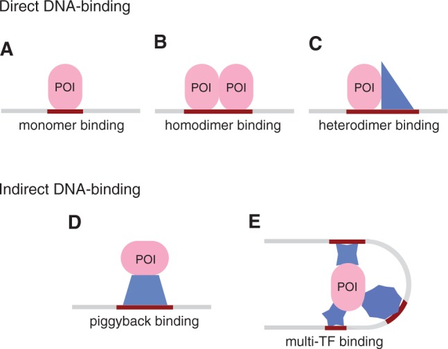
Figure 1.

Different modes of protein–DNA binding. The profiled protein is shown as an oval and co-factors as polygons. A direct DNA-binding protein can recognize different sites based on its partner: (A) a half-site as a monomer, (B) a symmetric motif as a homodimer, and (C) two different half-sites as a heterodimer. An indirect DNA-binding protein can immunoprecipitate sequences containing the consensus of (D) one or (E) several co-factors. See Farnham (6) for a discussion on why regions arising from ChIP experiments may not contain a match to the consensus motif.
