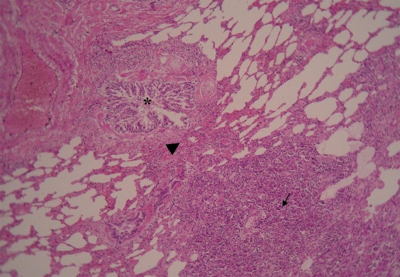
Figure 3.
Photomicrograph of a lung from a 12-year-old dog infected by Mycobacterium tuberculosis, showing granulomatous reaction with central necrosis, presence of epithelioid cells and fibroblasts in the middle zone (little arrow). Note proximity of granuloma (large arrow) with bronchial tree (*) indicating active tuberculosis (TB) infection (H&E stain, ×200).

