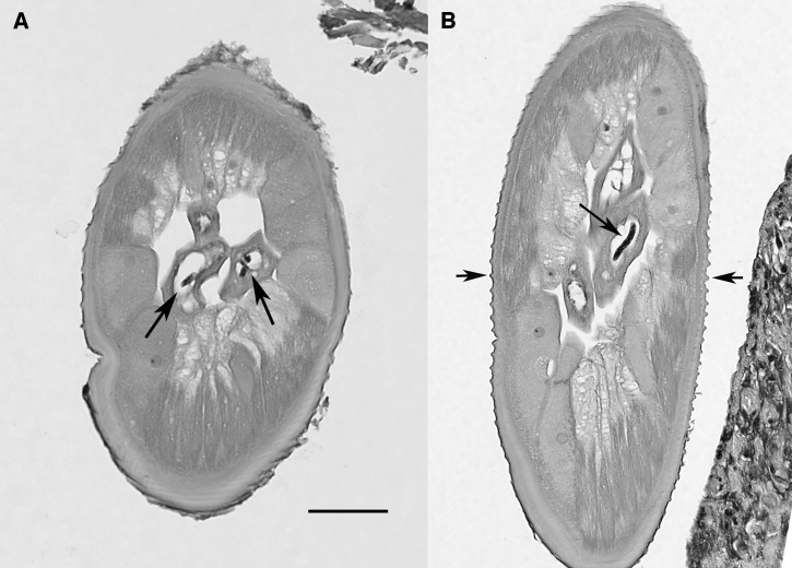
Figure 2.
Tissue sections of female Onchocerca lupi, hematoxylin and eosin stain. (A) Cross-section illustrating the morphology of the worm, including the detail of the muscle cells and large lateral chords, and the presence of microfilariae in utero (arrows). Scale bar = 50 μm. (B) Tangential section in which the same morphologic features are evident, including the presence of a microfilaria in utero (large arrow), and illustrating the distinctive cuticular ridges (small arrows). Scale bar same as in (A).

