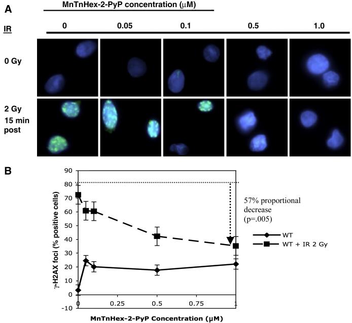
Figure 2.
Gamma-H2AX immunofluorescence foci formation in WT cells. WT cells were incubated in MnTnHex-2-PyP for 18 hours, were irradiated with 2 Gy and fixed with paraformaldehyde onto coverslips. After permeabilization with 0.5% Triton-X 100, cells were stained for γ-H2AX and cells were scored for presence of IRIF nuclear foci. (A) IRIF results for WT cells treated with MnTnHex-2-PyP. Note reduction in IRIF-positive cells with increasing concentrations of MnTnHex-2-PyP. (B) Quantitation of the IRIF results. Solid line: unirradiated WT cells; dashed line: 15 minutes post-2 Gy WT cells. The dotted guideline indicates the percentage of irradiated WT cells positive for IRIFs, in the absence of any compound; the dashed arrow indicates the maximal reduction in IRIF-positive cells after exposure to 1 μM MnTnHex-2-PyP. Error bars represent the standard error. All slides were coded by one individual and read blindly by another. The results shown here are the average results for 3 experiments.

