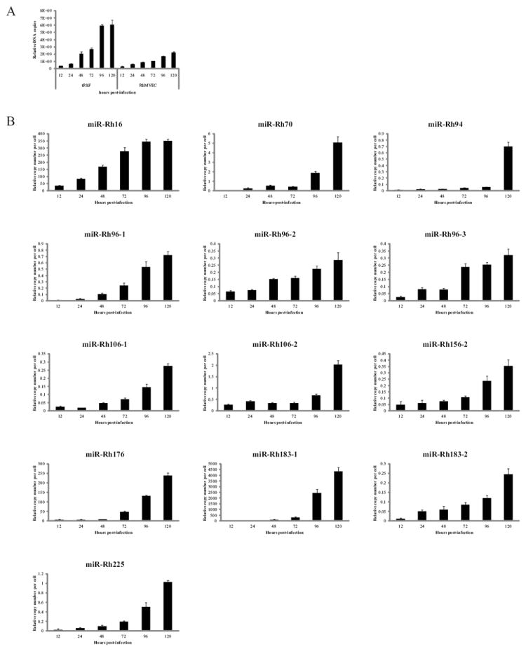
Figure 4. RhCMV miRNA expression in endothelial cells.

Rhesus microvascular endothelial cell (RhMVEC) or telomerized rhesus fibroblasts (tRhF) were mock-infected or infected with 2 PFU/cell of RhCMV and DNA and RNA was harvested at the indicated timepoints. (A) 100ng of DNA was used to perform Real-time PCR for Rh87. Data is presented as relative copy number as determined using serial dilution of RhCMV BAC DNA. Data was normalized to cellular β-actin. (B) Experiments were performed as in the legend of Figure 3.
