
Fig. 2.
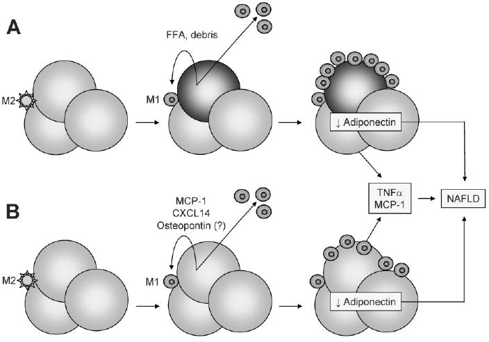
Proposed mechanisms of adipose tissue macrophage activation and their contribution to NAFLD. (A) Normal adipose tissue contains a small number of resident macrophages with an M2 (anti-inflammatory) phenotype. Expansion of adipocytes with fat in obesity can provoke adipocyte necrosis, with the released cellular debris and free fatty acids (FFA) activating resident macrophages and signaling the recruitment of M1 (proinflammatory) macrophages from the circulation. The resulting inflamed fat produces high levels of TNFα and MCP-1 and low levels of adiponectin, which can contribute to NAFLD. TNFα and MCP-1 can derive from both adipocytes and macrophages, whereas adiponectin is produced exclusively by adipocytes. (B) Obese adipocytes remain viable but are induced to secrete MCP-1, CXCL14, and perhaps osteopontin. This attracts and activates macrophages to an M1 phenotype. The end result is the same, with inflamed fat producing high levels of TNFα and MCP-1 and low levels of adiponectin.