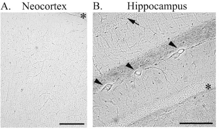
Figure 2.
Demonstrating cerebral vasculature in unstained sections from mouse neocortex (A) and hippocampus (B). Asterisk in A refers to pial surface. Arrow and asterisk in B refer to the CA1 pyramidal cell and dentate gyrus granule cell layers, respectively. Arrowheads in B point to large vessels at the hippocampal fissure. Scalebars in A & B: 250μm.

