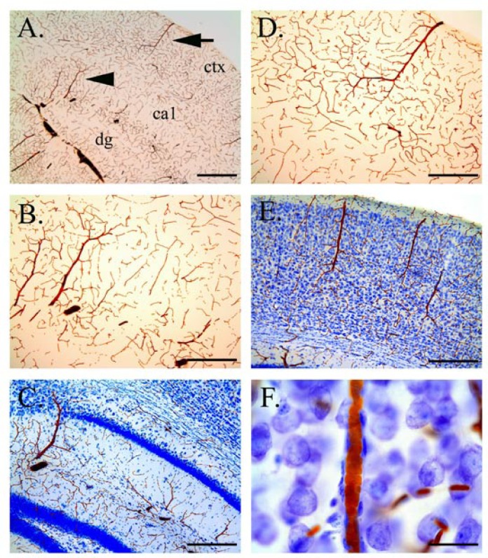
Figure 4.
Peroxidase staining of RBCs in unperfused sections. Arrowhead and arrow in A refer to vessels shown at higher magnification in B and D, respectively. Nissl-counterstained sections shown in C (hippocampus), E and F (neocortex). High magnification of stained RBS in a vessel and associated perivascular cells shown in F. Scalebars in microns: A, 500; B–E, 250, F, 25.

