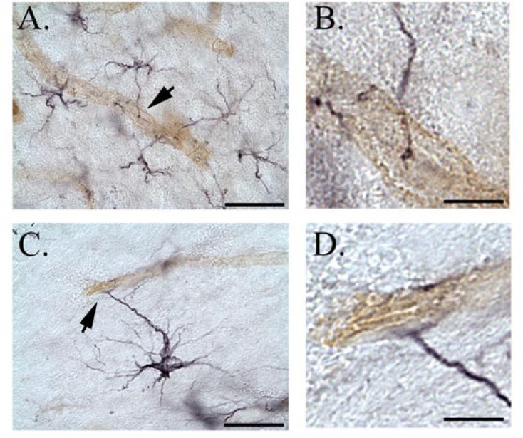
Figure 7.
Double immunocytochemistry against astrocyte-specific antigen GFAP (black staining) and the rat endothelial cell-specific antigen RECA (brown staining). A–D, Representative micrographs of rat neocortex. GFAP endfeet in A and C (arrows) are shown at higher magnification in B and D, respectively. Scalebars in microns: A, 250; C, 25; B and D 6.25.

