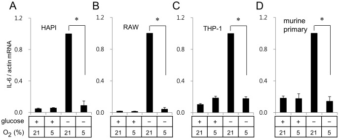
Figure 2. Hypoxia suppressed glucose deprivation-induced IL-6 expression in various types of cells.
Rat microglial cell line HAPI cells (A), murine macrophage RAW264.7 cells (B), human monocyte THP-1 cells (C), and murine primary microglia (D) were incubated for 7 h as described in Fig. 1A. Expression of IL-6 was analyzed as described in Fig. 1A. *p<0.01 vs glucose deprivation at 21% oxygen.

