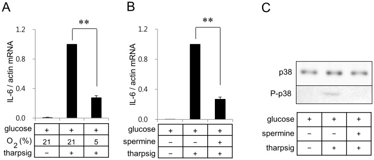
Figure 5. Hypoxia and spermine suppress thapsigargin-induced IL-6 gene expression in BV2 cells.
Real-time RT-PCR of IL-6 was carried out with RNA extracted from BV2 microglia incubated in a glucose containing serum free DMEM under the normoxic or hypoxic conditions (A). Real-time RT-PCR of IL-6 was carried out with RNA extracted from BV2 microglia incubated in a glucose containing serum free DMEM with or without 1.5 mM spermine (B). Thapsigargin (20 nM) or spermine were added as indicated. Results are shown as means ± SD; n = 3 independent experiments. **p<0.01. (C) Whole cell lysates were separated on SDS-polyacrylamide gels and analyzed with antibodies against p38 or P-p38. Quantitation of protein levels of P-p38 were carried out as described in Fig. 4C. *p<0.05.

