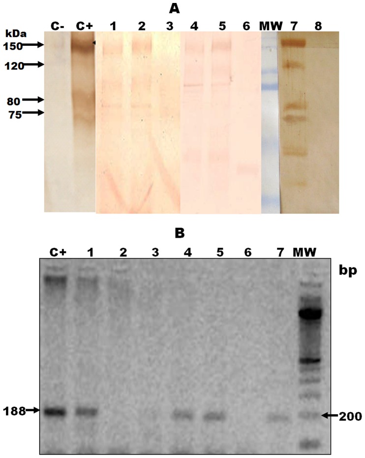
Figure 1. Detection of antigens and DNA of T. cruzi in urine of guinea pigs experimentally infected.
1.A. Antigenic bands in urine samples of guinea pigs infected with T. cruzi. Bands were detected by Western Blot using a polyclonal antibody against excretory-secretory trypomastigote T. cruzi antigen (TESA). C-: Negative control (RPMI 1640 medium). C+: Positive control (TESA antigen). MW: molecular weight marker. Urine samples of infected guinea pigs: Lane 1) 165 dpi, lane 2) 25 dpi, lane 4) 115 dpi, and lane 5 and 7) 55 dpi. Urine samples of non- infected guinea pigs: Lanes 3, 6 and 8. Bands under 70 kDa were considered unspecific because 25% of the non-infected guinea pigs had a reaction to these low bands. 1. B. Detection of trans-renal DNA in urine samples of guinea pig infected with T. cruzi. Bands were detected by PCR using primers TcZ1/TcZ2. C+: Positive control (DNA of T. cruzi from medium culture). MW: molecular weight marker. Urine samples of infected guinea pigs: Lane 1) 25 dpi, lane 3) 55 dpi, lane 4) 40 dpi, lane 5) 55 dpi, and lane 7) 25 dpi. Urine samples of non- infected guinea pigs: Lanes 2 and 6.

