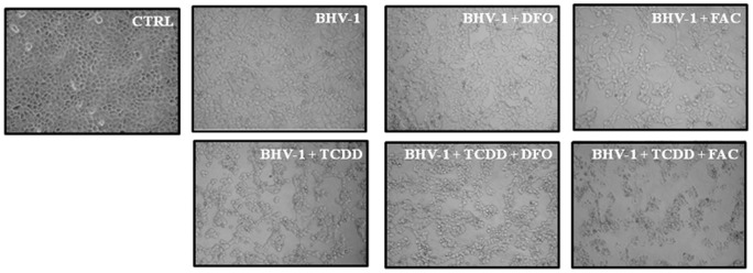
Figure 6. Effect of iron repletion-depletion on CPE.

Representative microphotographs by phase-contrast light microscopy of iron deplete-replete MDBK cells exposed or not, as indicated in the legends, to 1 pg/ml of TCDD at 48 h post-BHV-1 infection (MOI of 1), showing the cytopathic effects and the morphological changes on cellular monolayers. In iron depletion – repletion experiments, cells were treated with 100 μM desferrioxamine (DFO) or with 50 μg/ml ferric ammonium citrate (FAC).
