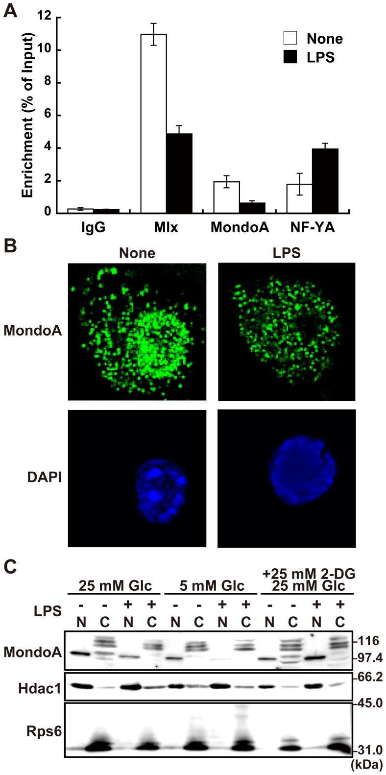
Figure 4. Dissociation of MondoA:Mlx from the Txnip promoter on LPS stimulation.
(A) Chromatin immunoprecipitation assay was performed to examine occupation of Mlx, MondoA, and NF-YA in Txnip promoter region with RAW264.7 cells stimulated with or without 100 ng/ml LPS for 45 min. Enriched DNAs was eluted and Txnip promoter region was quantified by quantitative PCR. Data shown are mean ± S.E. of duplicate samples. (B) RAW264.7 cells stimulated with or without 100 ng/ml LPS for 45 min. Cells were fixed, permeabilized, and stained with anti-MondoA antibody and Alexa488-conjugated polyclonal anti-rabbit IgG antibody. Fluorescence microscopic images were obtained and analyzed by three-dimentional deconvolution. (C) Nuclear and cytoplasmic fractions of RAW264.7 cells stimulated with or without 100 ng/ml LPS for 45 min were analyzed by western blotting with anti-MondoA, histone deacetylase 1 (Hdac1) or ribosomal S6 ribosomal protein (Rps6). Data shown are a representative of at least three independent experiments.

