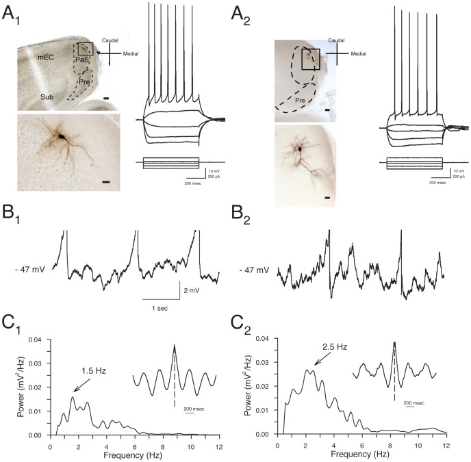
Figure 1. Layer II parasubucular (PaS) stellate and pyramidal neurons show similar electrophysiological properties.
A. Photomicrographs of representative layer II stellate cell (A1) and pyramidal cells (A2) show cell bodies in layer II near the border with layer I. Calibration bars are 100 µm in top panels, and 20 µm in bottom panels. Membrane potential responses to hyperpolarizing and depolarizing current pulses for the cells shown were similar for stellate and pyramidal neurons (right panels). B. Both stellate (B1) and pyramidal (B2) neurons display theta-frequency membrane potential oscillations at near-threshold voltages. Representative traces obtained from the same cells as in A show oscillations in membrane potential when cells were depolarized to −47 mV using positive current injection. Note that action potentials are truncated. C. Power spectra reflect a peak frequency of oscillations at 1.5 to 5.9 Hz in these neurons recorded at 22–24 °C. The rhythmicity of oscillations is also reflected in the inset autocorrelograms.

