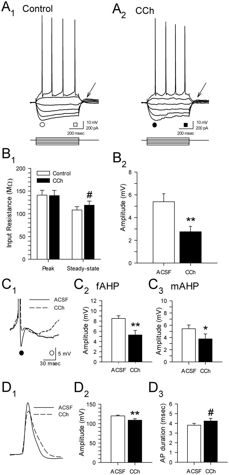
Figure 3. Carbachol has multiple effects on electrophysiological properties of layer II parasubicular neurons.
A. Membrane voltage responses to hyperpolarizing and depolarizing current pulses in the same cell during perfusion with control ACSF (A1) and 25 µM CCh (A2). B. Group data in B1 show that CCh (25 µM) resulted in an increase in steady-state input resistance (measured at times indicated by squares in A; *: p<0.05), but had no effect on peak input resistance (see circles in A). Carbachol consistently reduced the amplitude of anodal break potentials following −200 pA steps (B2, **: p<0.01; see arrows in A). C. Magnified superimposed traces (C1) in control ACSF (solid line) and in the presence of 25 µM carbachol (dashed line) show a reduction in the average amplitude of the fast (C2; •, fAHP, **: p<0.01) and the medium afterhyperpolarization (C3; ○, mAHP, *: p<0.05). D. Superimposed action potentials show that carbachol (25 µM) was also associated with a significant reduction in action potential amplitude (C1,2; **: p<0.01) and a trend towards increased spike duration (C3; #: p = 0.06).

