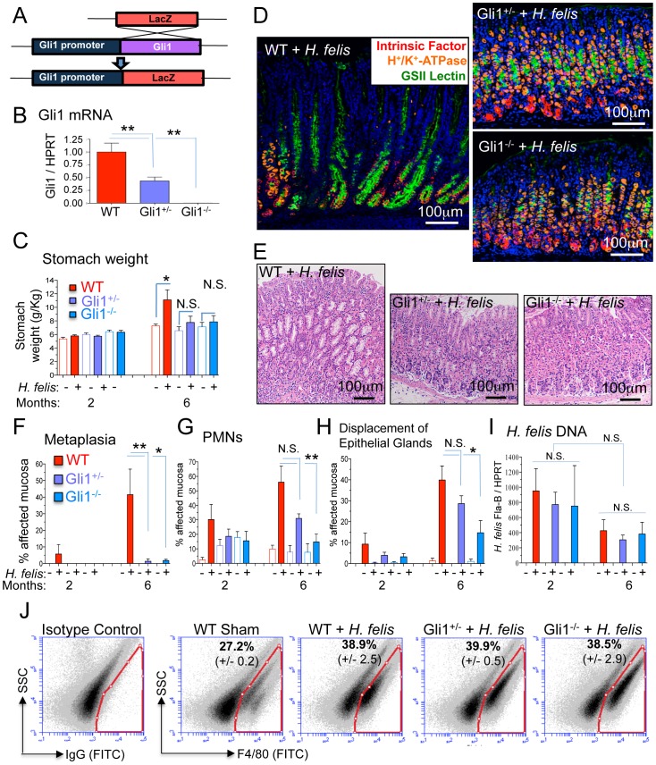
Figure 1. Gli1 deletion prevents gastric metaplasia.
A) Schematic representation of LacZ substitution for the Gli1 gene to generate Gli1+/− and Gli1−/− mice. B) Gli1 mRNA in stomachs of WT, Gli1+/− and Gli1−/− mice. C) Stomach weight normalized to total body weight in infected and uninfected mice. D) Triple immunofluorescent staining of Griffonia simplicifolia II (GSII) lectin (green; mucous neck cells), intrinsic factor (red; chief cells), and H+/K+-ATPase (orange; parietal cells) in 6-month H. felis-infected WT, Gli1+/− and Gli1−/− stomachs. E) Hematoxylin and eosin (H&E) staining of the gastric mucosa in 6-month H. felis-infected WT, Gli1+/− and Gli1−/−. F–H) Histologic scoring of metaplasia, PMN infiltration, and displacement of epithelial glands by infiltrating inflammatory cells in 2- and 6-month H. felis-infected WT, Gli1+/− and Gli1−/− stomachs. I) qPCR quantification of H. felis flagellar filament B (Fla-B) DNA in the infected mucosa. J) Flow cytometric analysis of F4/80+ cells versus side-scatter in WT Sham and 6-month infected WT, Gli1+/− and Gli1−/− stomachs (N = 3 mice per group). Open and closed bars denote uninfected and infected mice respectively. For RT-qPCR experiments, N = 5–10 mice per group. Error bars represent the mean +/− SEM. ***p<0.001; **p<0.01; *p<0.05. N.S. = not significant.

