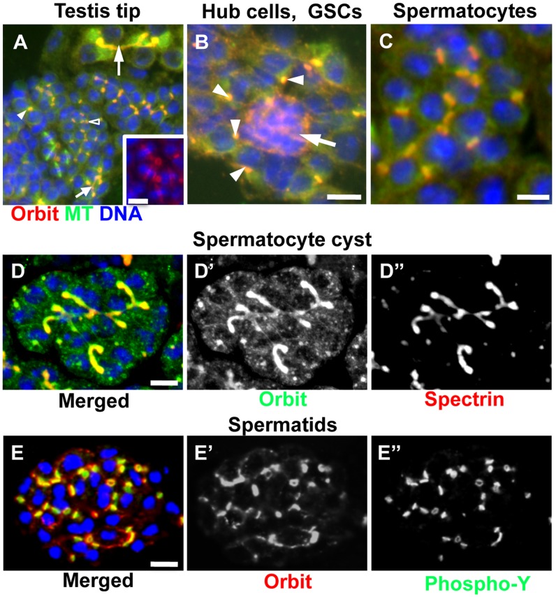
Figure 1. Orbit immunostaining and expression of Orbit fused with fluorescence tags in testis tip cells.
(A) Lower magnification view of cells derived from testis tip. Microtubules (green), anti-Orbit immunostaining (red), and DNA (blue) are shown. Orbit is localized on a spectrosome in a 2-cell cyst (smaller filled arrowhead), and on a fusome in an 8-cell cyst (smaller filled arrow). It is also localized on ring canals in a 16-cell cyst (open arrowhead). The larger arrow shows Orbit localization on pieces of fusome formed in a partial spermatocyte cyst at the S2b stage, i.e., the early stage of the growth phase. Inset; a 4-cell spermatogonial cyst at metaphase. (B) Hub cells (arrow) surrounded by several germline stem cells (GSCs). Orbit (red) is highly concentrated in the cytoplasm of hub cells, and on single spectrosomes (arrowheads) in the GSCs. (C) Orbit immunolocalization on ring canals in an early spermatocyte cyst. (D) Expression of GFP-Orbit in an early spermatocyte cyst at the S3 stage, i.e., the middle of the growth phase (green in D, D′). Anti-spectrin immunostaining (red in D, D″) to visualize the fusome is shown. (E) Expression of mRFP-Orbit in a partial spermatid cyst at the onion stage (red in E, E′). Anti-phospho-tyrosine immunostaining (green in E, E″) to visualize ring canals is indicated. Scale bar = 10 µm.

