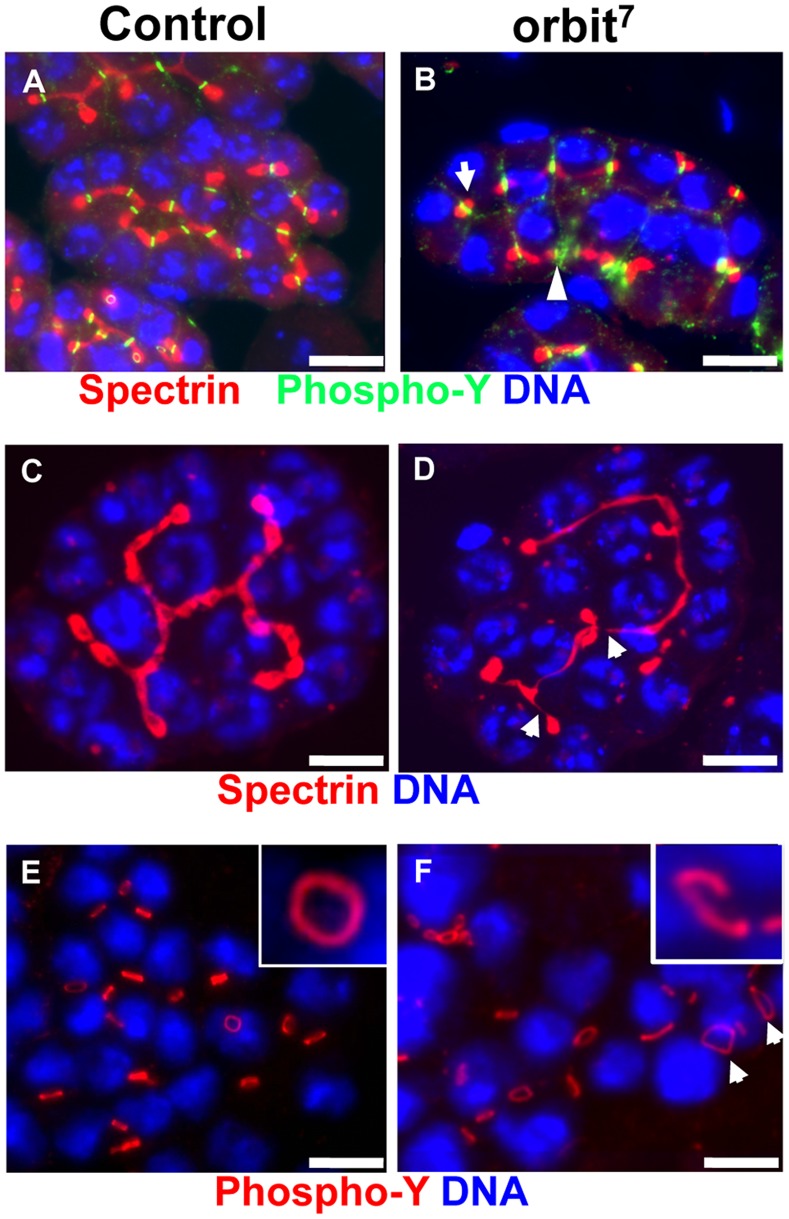
Figure 5. The orbit gene is required for development of fusomes and ring canals in spermatocyte cysts.
(A, B) Immunostaining of early spermatocyte cysts from normal males and from orbit7 mutant males by using anti-α-spectrin antibody for fusome visualization (red) and anti-phospho-tyrosine for ring canal observation (green). DNA staining (blue). Note the abnormal fusomes, which failed to elongate (arrow) or branch in the mutant cysts. The ring canal marker failed to be incorporated in the lumen of ring canals (arrowhead). (C) A normal branched fusome structure with constant thickness in a wild-type early spermatocyte cyst at the S2 stage. (D) A less-branched fusome structure in the mutant spermatocyte cyst. The abnormal fusome becomes thinner in places or disconnected (arrows). (E) Immunodetection of ring canals in early spermatocyte cysts using anti- phospho-tyrosine antibody. Note the formation of ring canals with constant diameter between every nucleus (blue) in wild-type spermatocytes. Note also that normal ring canals are shaped by a continuous hollow structure (inset). (F) In early spermatocytes from orbit7 mutant males, disconnected ring canals are observed in the mutant cysts (inset). Abnormal ring canals with larger diameters (arrows) are observed in early spermatocytes from orbit7 mutant males. Scale bar = 10 µm.

