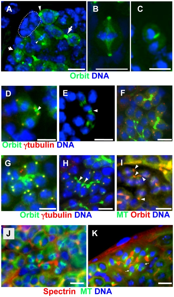
Figure 6. Dynamic alteration of Orbit localization in germline stem cells (GSCs) and spermatogonial division.
(A–H) GFP-Orbit (green) and DNA (blue) are shown. (D, F, G, H) anti-γ-tubulin immunostaining (red). (I) GFP-tubulin (green), anti-Orbit immunostaining (red), and DNA (blue). (J, K) GFP-tubulin (green), anti-spectrin antibody immunostaining (red), and DNA (blue). (A) Tip of a testis from a nos-Gal4>UAS-GFP-Orbit male. Hub cells are encircled by a dotted line. Orbit is localized on the spectrosome of a GSC (larger arrowhead), kinetochores of a GSC at metaphase (smaller arrowhead), central spindle microtubules of a presumptive GSC (smaller arrow), and fusome plug formed at the midbody of a presumptive GSC (larger arrow). (B) A gonialblast undergoing the first division at metaphase. (C) Late anaphase of a gonialblast. (D) In the second spermatogonial division, two centrosomes (red) are connected by a single spectrosome (arrowhead). Orbit is more abundant on spectrosomes and around centrosomes attached to the spectrosome, than on distal centrosomes. (E) A 2-cell cyst of dividing spermatogonia at anaphase. Note the contractile ring (arrowhead) between two cells. (F) Two 4-cell cysts and part of a 16-cell cyst at interphase. Most of the centrosomes (red) are not associated with fusomes at interphase. (G) An 8-cell cyst in which γ-tubulin foci (yellow) have become evident at the onset of the third mitotic division. (H) Prophase to prometaphase of the third spermatogonial division. Every centrosome pair (red) orients towards the fusome, and one of the two centrosomes (arrowheads) is captured by the fusome. (I) Immunostaining of an 8-cell cyst at metaphase. Orbit is localized on kinetochores and part of the fusome (arrowheads). (J, K) An 8-cell spermatogonial cyst undergoing mitosis; (J) normal control male and (K) orbit7 mutant male. The mutant fusome appears to be disconnected. At least two mitotic spindles (arrows) have been detached from the fusome.

