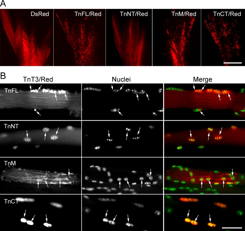
Fig. 5.
Subcellular localization of TnT3/DsRed proteins in mouse FDB fibers. TnT3/DsRed constructs and control plasmids were electroporated in vivo into the mouse FDB muscle. a Expression pattern of all constructs in whole isolated muscle. Control DsRed and TnNT/DsRed were expressed evenly throughout the muscle. In contrast, TnFL/DsRed, TnM/DsRed, and TnCT/DsRed showed a punctate distribution. b Higher magnification imaging analysis of individual myofibers showed that both TnFL/DsRed and TnCT/DsRed localized in some myonuclei. Additionally, TnFL/DsRed showed weak striated pattern. Notably, these two constructs localized at different subnuclear domains as revealed by their different co-localization pattern with Hoechst 33342 DNA staining. TnNT/DsRed also expressed in the myonuclei, but the myonuclear DNA staining pattern was not affected. TnM/DsRed mainly localized in the cytoplasm as small puncta, while most of it was highly ordered and reflects binding to myofibrils. Arrows point to nuclei in red fluorescence-positive areas. Scale bars, 800 μm (a), 50 μm (b). Fibers are representative of at least 200 fibers in each group from 2 to 13 experiments (DsRed, n = 3; TnFL/DsRed, n = 5; TnNT/DsRed, n = 6; TnM/DsRed, n = 2; TnCT/DsRed, n = 13)

