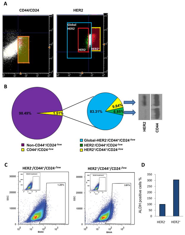
Figure 2.
Characterization of the HER2+/CD44+/CD24−/low BCSCs within MCF7/C6 cell lines. (A) CD44+/CD24−/low cells were sorted from 2.5 × 10⁷ MCF/C6 cells with CD44 and CD24 antibodies (left panel), from which the cells were further sorted with HER2 antibody (right panel). The orange box in the left panel indicates the CD44+/CD24−/low fraction used for sorting HER2+/CD44+/CD24−/low (yellow box), HER2−/CD44+/CD24−/low cells (red box) and a global HER2−/CD44+/CD24−/low population (blue box with a broader gate). (B) Percentages of CD44+/CD24−/low and HER2+/CD44+/CD24−/low cells derived from MCF7/C6 cells (left panel, also shown in Table S2). Western blot analysis of HER2 and CD44 protein expression in sorted HER2+/CD44+/CD24−/low and HER2−/CD44+/CD24−/low cells (right panel). (C) Analysis of ALDH activity in HER2−/CD44+/CD24−/low versus HER2+/CD44+/CD24−/low cells via flow cytometry using Aldefluor staining. As negative control, cells were incubated with ALDH inhibitor DEAB. (D) Quantification of the ALDH expression HER2+/CD44+/CD24−/low compared to HER2−/CD44+/CD24−/low BCSCs analysis in C.

