
Fig. 2.
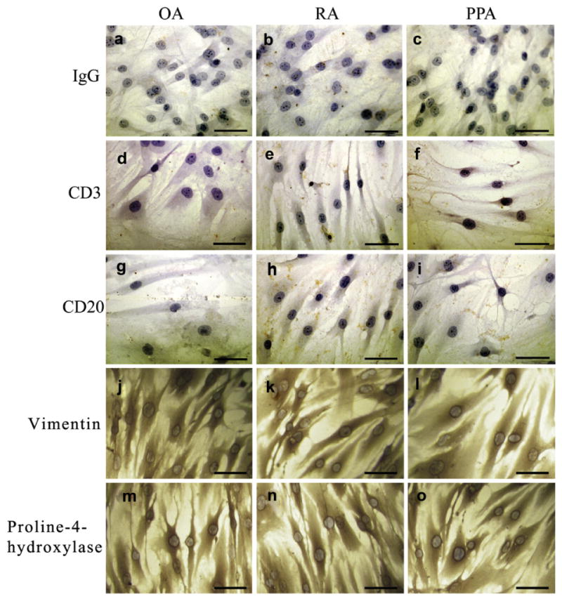
Immunohistochemistry of isolated synovial fibroblasts. Fibroblasts cultured on coverslips showing the (a–c) IgG control and lack of expression of (d–f) the T cell marker CD3 and (g–i) the B cell marker CD20 and positive expression of (j–l) vimentin and (m–o) proline-4-hydroxylase. Scale bar represents 50 μm.