
Fig. 4.
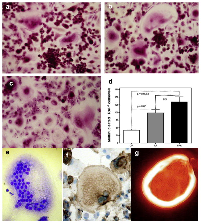
TRAP cytochemical stain images of human PBMC cultured for 16 days in the presence of M-CSF and conditioned medium from a) OA, b) RA and c) PPA and results expressed d) graphically, (p < 0.05 Manne–Whitney nonparametric test, error bars standard error of the mean) data pooled from 3 experiments. Osteoclasts were also determined by the presence of multinucleated cells (e) expressing the specific osteoclast markers (f) VNR, and capable of (g) F-actin ring formation. (Representative pictures of at least three experiments M-CSF and CM OA, RA and PPA cultures that induced osteoclast formation).