
Fig. 5.
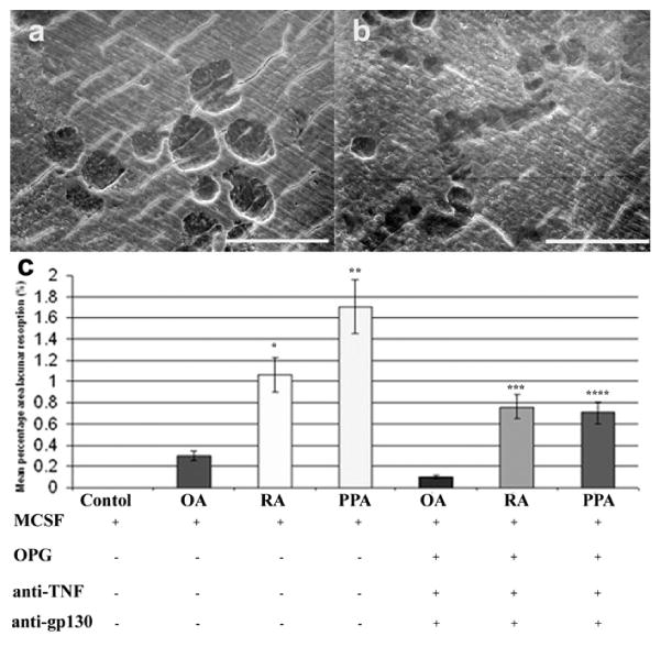
Scanning electron micrographs of dentine slices after PBMC culture for 21 days in the presence of M-CSF, OPG, and CM of cultured arthritic synovial fibroblasts. Fibroblasts isolated from (a) OA and (b) RA showed evidence of lacunar resorption formation. (Image representative of average lacunar resorption formation.) (c) Mean percentage area of lacunar resorption in OA, RA, and PPA conditioned medium in cultures of PBMCs incubated for 21 days in the presence of M-CSF, with or without OPG, anti-TNF, and anti-gp130. Scale bars represent 100 μm. (Representative data of at least three experiments.)