
Figure 6.
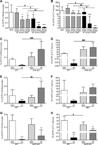
Infection with M. avium 25291 induces depletion of the early thymocyte precursors and depends on IFNγ production, IFNγ-dependent macrophage activation and NO production.
A, B. Percentage (A) and number (B) of ETP from non-infected (Ninf) thymi and from thymi of WT mice infected with M. avium strains 2447 or 25291. C, D. Percentage (C) and number (D) of ETP from non-infected (Ninf) thymi and from thymi of WT or IFNγ-KO mice infected with M. avium 25291 at 80 dpi. E, F. Percentage (E) and number (F) of ETP from non-infected (Ninf) thymi and from thymi of WT or MIIG mice infected with M. avium 25291 at 80 dpi. G, H. Percentage (G) and number (H) of ETP from non-infected (Ninf) thymi and from thymi of WT or iNOS-KO mice infected with M. avium 25291 at 80 dpi. Data represent the mean of the percentages or the mean and standard deviation of the number of cells. Each group of mice comprised three to six animals analysed individually. Data shown represents one experiment out of three. Statistically significant differences between infected and non-infected control mice are labeled as *p<0.05, **p<0.01, ***p<0.001. Statistically significant differences between infected groups are labeled as #p<0.05, ##p<0.01, ###p<0.001.