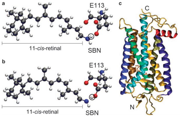
Figure 1.
The 11-cis-retinal and a visual pigment. (a) Protonated Schiff base nitrogen-linked 11-cis-retinal. The circled hydrogen atom (in white) is bound to the Schiff base nitrogen. (b) Unprotonated analog, where the circled hydrogen atom is bound to the carboxylic oxygen of E113 of the visual pigment. The black, blue, red, and white colors represent carbon, nitrogen, oxygen, and hydrogen atoms, respectively. (c) The seven transmembrane helices and 11-cis-retinal of bovine rhodopsin (pdb 1U19). The 11-cis-retinal is shown with the ball and stick model near the center, and N and C indicate the amino and carboxyl termini of the visual pigment.

