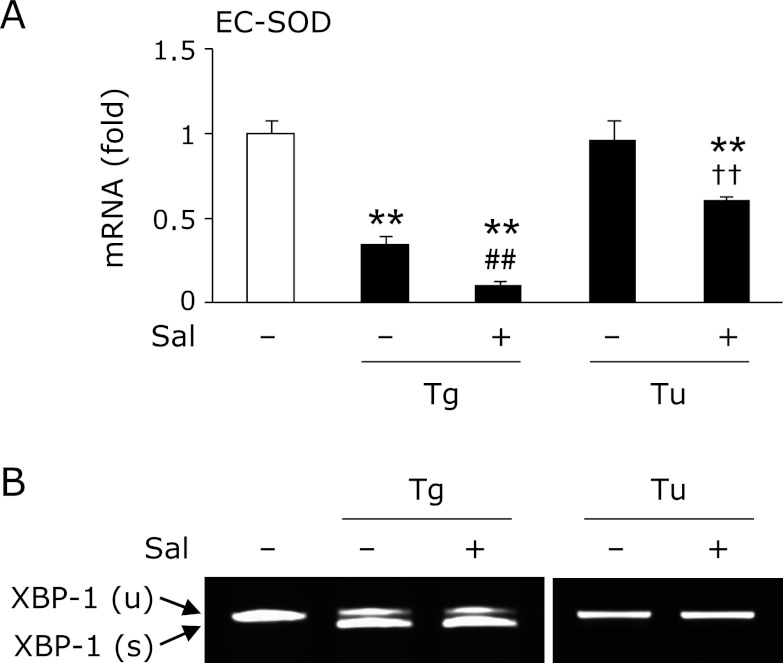
Fig. 4.
Involvement of eIF2α signaling in the thapsigargin-triggered reduction of EC-SOD. 3T3-L1 adipocytes were pretreated with (+) or without (−) 15 µM salubrinal (Sal) for 1 h, and then treated with 1 µM thapsigargin (Tg) or 1 µg/mL tunicamycin (Tu) for 24 h. After the cells had been treated, RT-PCR (A, B) was carried out. RT-PCR data (A) were normalized using GAPDH levels (**p<0.01 vs vehicle, ##p<0.01 vs Tg-treated cells, ††p<0.01 vs Tu-treated cells).

