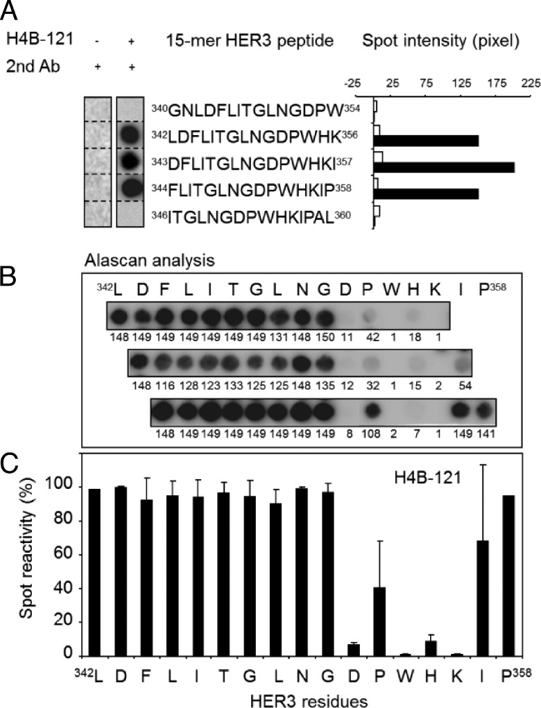
Figure 2.
The 352DPWHKI358 motif recognized by the Ab H4B-121 is located in D3 of HER3. (A) Scan analysis of the 213 overlapping pentadecapeptides that covered the entire extracellular domain of HER3 (amino acids 1–645) and were frame-shifted by one or three residues (generated by the SPOT method). Binding of H4B-121 was observed only in the region between amino acids 340 and 360. No binding was observed with the secondary Ab alone. The Spot intensity was measured using ImageJ. (B) Alanine scanning (Alascan) of the pentadecapeptides 342LDFLITGLNGDPWHK356, 343DFLITGLNGDPWHKI357, and 344FLITGLNGDPWHKIP358 that cover the region between amino acids 342 and 358 of HER3. (C) Quantitative analysis of H4B-121 binding to the membrane pentadeca-peptides in B using ImageJ. Each bar represents the reactivity of H4B-121 toward a pentadecapeptide sequence inwhich the indicated amino acid was substituted by alanine. The mean spot reactivity for each residue was calculated from the results obtained in the three pentadecapeptides.

