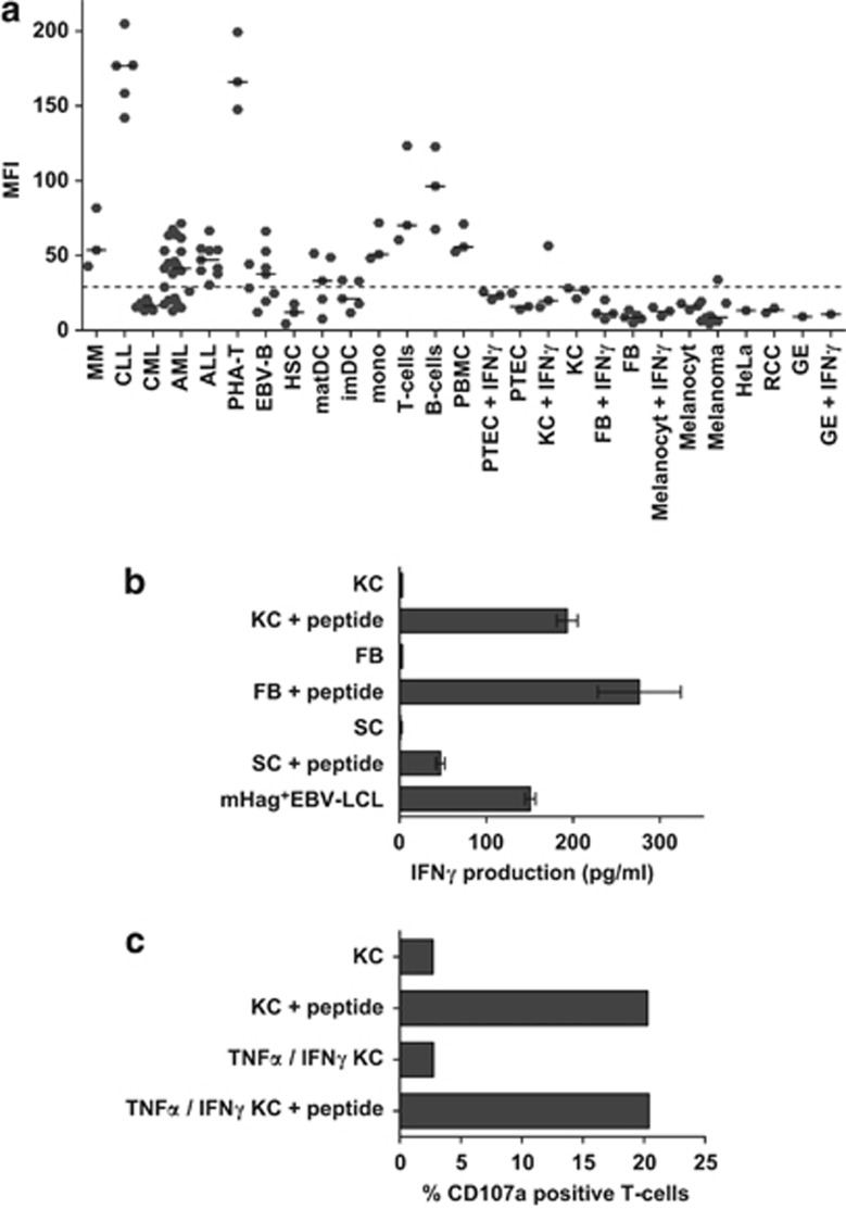
Figure 3.
UTA2-1 expression is restricted to hematopoietic tissue and CTL 503A1 can lyse various malignant hematopoietic mHag+ cells. (a) The expression level of gene C12orf35 was assessed in an established microarray, showing an overexpression of the gene in hematopoietic tissue and low expression levels in non-hematopoietic cells. Among the hematopoietic cells, expression is highest in CLL cells and PHA T-cell blasts,and lowest in hematopoietic stem cells, immature dendritic cells and CML cells. We set the cutoff for T-cell recognition on a MFI of 30 according to our findings in functional experiments using EBV-LCL and keratinocytes. (b) The non-hematopoietic cell types fibroblasts, keratinocytes and stromal cells were not recognized by CTL 503A1 in an E:T ratio of 5:1 using IFN-γ ELISA. After incubating the cells with 10 μℳ exogenous peptide, 503A1 did become activated. (c) The keratinocytes did not induce an increase in expression of degranulation marker CD107a on CTL 503A1 in a E:T ratio of 5:1 as measured by FACS. This was independent of pre-incubation with tumor necrosis factor-α (TNF-α) and IFN-γ (both 100 IU/ml) for 3 days. After incubating the target cells with 10 μℳ exogenous peptide, CD107a on the CTL was upregulated.

