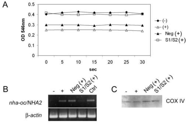
Figure 7.
NHA-oc/NHA2 mediates Na+- induced mitochondrial swelling.
(A) RAW 264.7 cells were transfected with a mix of nha-oc/NHA2-specific siRNA molecules and stimulated with RANKL. 4 days later mitochondria were isolated and resuspended in Na+-swelling buffer. For this experiments we used equivalent amounts of mitochondria (100μg protein). OD546 was recorded every 5 sec for 30 sec. A drop in OD546 indicates swelling of the mitochondrial matrix. (-) unstimulated cell; (+) RANKL stimulated; (Neg) RANKL stimulated, treated with a control siRNA; (S1/S2) RANKL stimulated, treated with a specific siRNA mix.
(B) RNA from RAW264.7 cells, was analyzed by RT-PCR using nha-oc/NHA2-specific (top) or p-actin (bottom) primers. (-) un-stimulated cells; (+) RANKL stimulated; (Neg) RANKL stimulated, treated with a control siRNA; (S1/S2) RANKL stimulated, treated with a specific siRNA mix. Densitometric analysis of the gel shows that S1/S2 treatment results in a reduction of >90% in the nha-oc/NHA2 amplification product.
(C) As a loading control, 10 μl of each mitochondrial fraction were analyzed by Western Blot analysis using a mitochondrial-specific COX IV antibody. A single 18KDa band of equal intensity can be seen in all lanes.

