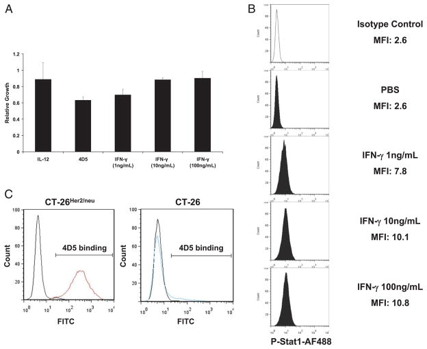
FIGURE 4.
IFN-γ does not inhibit proliferation of CT-26HER2/neu cells. A, CT-26HER2/neu cells were treated in vitro with IL-12, trastuzumab, or increasing concentrations of IFN-γ. Proliferation was determined by the MTT assay. B, CT-26HER2/neu cells were stimulated for 15 min in vitro with increasing concentrations of IFN-γ or PBS. The percentage of cells containing activated STAT1 was assessed by flow cytometry. The x-axis of each histogram represents the specific fluorescence of p-STAT1 on a four-decade logarithmic scale, and the y-axis represents the total number of events. C, CT-26HER2/neu cells and parental CT-26 cells were analyzed for the binding of 4D5 to cell surface HER2/neu by flow cytometry using the 4D5 Ab and an FITC-labeled rabbit anti-murine Ab.

