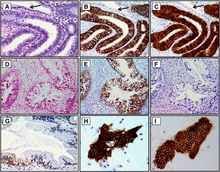
Figure 2.
Examples of CA-IX and p16 immunoreactivity in CGL, GA and CA-IX expression in normal cervix and exfoliated endocervical cells. Strong membranous reaction for CA-IX (B) and cytoplasmic and nuclear staining for p16 (C) was seen in all cases of CGL, but the normal gland was non-reactive for CA-IX or p16 (B and C, arrows). In contrast, in cases of GA only strongly positive CA-IX staining was seen (E), and p16 expression was either weakly cytoplasmic or negative (F). In normal cervical glands, CA-IX expression was limited to a few columnar/reserve cells, and the staining was weak and limited to the cytoplasm (G, arrow). Atypical glandular cells (H) derived from GA and normal looking glandular cells (I) derived from LEGH with atypia showed diffuse strong membranous positivity for CA-IX. The relevant haematoxylin and eosin (H&E)-stained tissue sections are shown in (A) and (D). Original magnifications: × 200 in (D–F) and × 400 in (A–C, G–I).

