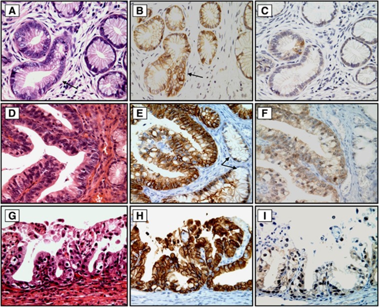
Figure 3.
Histology and immunohistochemical staining for CA-IX and p16 protein expression in LEGH with atypia. In contrast to Figure 1B, CA-IX immunoreactivity was not only limited to the central larger glands but was also observed in the atypical glands (B, arrow, and E). Diffuse CA-IX membranous positivity was also seen in LEGH with atypical papillary growth (H). Note that the small glands without atypia were CA-IX negative (E, arrow). In contrast, p16 expression in all cases of LEGH with atypia was either negative or exhibited very weak cytoplasmic staining (C, Fand I). The relevant haematoxylin and eosin (H&E)-stained tissue sections are shown in (A, D and G). Original magnification: × 400.

