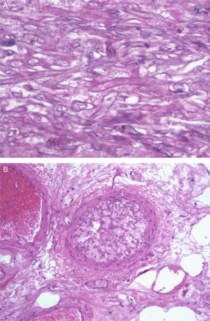
Fig. 1.
(A) Methenamine silver stain of the necrotic tissue at × 400 magnification within the surgical specimen revealed broad, branching, non-septated fungal elements characteristic of Rhizopus growth at 37°C. (B) Methenamine silver stain of the necrotic tissue at × 100 magnification shows a thrombosed arteriole infiltrated with non-septated fungal elements, establishing the diagnosis of angioinvasive Rhizopus.

